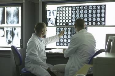
Medical imaging has revolutionized the evaluations of tissue anatomy and the functional performance of many organs. Yet many diseases such as cancer start at a molecular level, requiring molecular imaging assessments. Researchers at the University of Arizona are leading efforts to image the molecular compositions of tumors in patients with cancer, to improve cancer diagnoses and evaluate the earliest responses to cancer treatment.
Marty Pagel, PhD, and his colleagues of the Contrast Agent Molecular Engineering Laboratory (CAMEL) have developed innovative methods to measure the acid content in tumors. Just as our muscles “feel the burn” after exercise causes lactic acid to be produced, tumors are also very metabolically active and produce excess lactic acid. Their imaging method uses Magnetic Resonance Imaging (MRI) to identify acidic tumors with high metabolism, which is commonly used for noninvasively identifying tumors throughout the body.
 “We have been surprised to learn that some aggressive tumors in patients are more acidic than we anticipated,” said Dr. Pagel. “This supports other evidence that tumor acidosis is important in driving cancer progression.” In particular, lactic acid is associated with tumor growth and invasion into nearby tissues, and is also related to tumor metastasis to distant organs.
Furthermore, this molecular imaging method can detect a decrease in tumor acidity after starting drug treatment, which can provide a very early assessment of the drug’s effect on tumor metabolism. According to Christine Howison, Director of Pre-Clinical Imaging in CAMEL, “Our collaborations with pharmaceutical researchers at the University of Arizona and in industry have shown a remarkable change in tumor acidosis within one week of initiating treatment. Our eventual goal is to rapidly inform patients that the drug’s effect is working on the tumor, rather than waiting weeks and months to learn if the tumor is shrinking in size.”
This research program has required a highly multidisciplinary approach, involving researchers throughout the College of Medicine at the University of Arizona in Departments of Medical Imaging, Biomedical Engineering, Chemistry & Biochemistry, Pharmacy, Radiation Oncology, Obstetrics & Gynecology, and the University of Arizona Cancer Center. The Department of Medical Imaging has been particularly instrumental in establishing strong research collaboration with Siemens Medical Solutions to translate this molecular imaging method to clinical MRI instruments. For these many reasons, CAMEL has found an ideal location in the Sonoran Desert for their molecular imaging research at the University of Arizona.
Visit the UA Contrast Agent Molecular Engineering Laboratory (CAMEL)
Visit the UA Department of Medical Imaging


