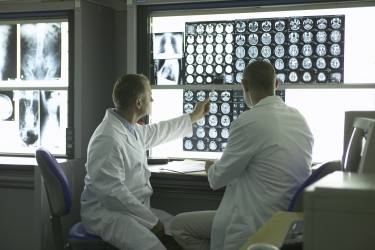
There has been a degree of convergence of the fields of radiology and pathology, with the term “radiomics” recently coined. Both fields are rapidly adapting to the digital age, and the tools being used in both fields are allowing detection and observation of smaller, more specific, and often personal features of disease. This is leading to increasingly individualized evaluation of organs, lesions, tissues, or even an ongoing dynamic molecular disease process.
This innovation is accelerating across the supply chain, from hardware design, to image acquisition, to post-image processing and analysis. Medical imaging is increasingly guiding interventions, often in real-time. Clinically annotated medical images, with genomic information are becoming readily available in research data repositories, ushering in the use of crowd-based analysis and advanced “big” data science to link phenotypes and genotypes in ways that have never been possible before.
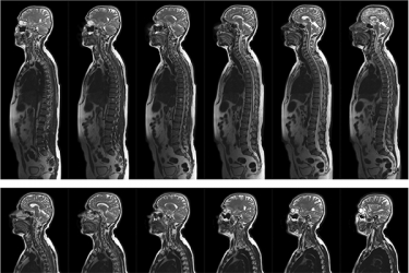
Researchers are using medical imaging in research to understand the commonalities and personal differences in disease at the population and individual levels. Collectively, new medical imaging technologies and their applications are allowing diagnosis and treatment from the molecular to the population level.
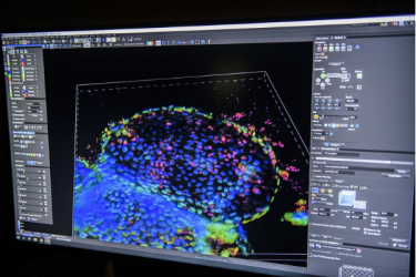
At the University of Arizona, the Department of Molecular Imaging as well as the Contrast Agent Molecular Engineering Laboratory (CAMEL) are developing new, innovative imaging techniques that can be used to identify and track disease progression and treatment.